Viability scan
For many women, pregnancy feels powerful and an amazing experience. After all, it makes her so happy and proud in making another human and bringing into this world. But for all its joyfulness, pregnancy can also be difficult and complex with problems that can occur throughout the gestation.
The most important aspect is the fear of losing the pregnancy (miscarriage) and the risk of this happening is higher during the first trimester. While it can be symptomatic in terms of bleeding and pain in most cases, it can also be asymptomatic (missed abortion or missed miscarriage) in a few.
Viability scan is performed between 6-10 weeks of pregnancy in women who have symptoms like bleeding or pain or in those who had previous history of miscarriages or ectopic pregnancies. The scan is performed either transabdominal or transvaginal to confirm viability and reassure the couple.
The scan takes approximately 15-30 minutes to perform and we would suggest a full bladder for the examination. A copy of the report would also be sent to the referring Doctor for further management.

Viability scan
Combined FTS (NT scan)with early anomaly assessment
It’s a general conception (rather misconception) that the incidence of chromosomal abnormalities increases with advance maternal age. Traditionally invasive testing like amniocentesis and CVS have been offered to pregnant women aged 35 years or more which can be associated with a risk of miscarriage following an interventional procedure.
Screening by a combination of maternal age, measurement of nuchal translucency and placental products of free Beta HcG and PAPP-A significantly improve the detection rate of common chromosomal abnormalities of up to 90%(5% FPR) thereby avoiding unnecessary fetal invasive procedures in a low risk patient. In addition, it also gives us an opportunity to perform an early anomaly assessment at this gestation to identify structural abnormalities that could be evident in the developing fetus at this stage.
The scan approximately takes about 30 - 45 minutes to perform, and it includes the following.
1. Measurement of the CRL (Crown to rump length) to calculate the estimated due date of delivery (EDD)
2. Measurement of nuchal translucency
3. Early fetal anomaly assessment
4. Maternal bloods to check levels of Beta HcG, PAPP-A to give a combined risk assessment for the common chromosomal abnormalities (e.g. Down syndrome)
5. Cervical assessment to predict risk of preterm delivery
Pregnant mothers undergoing this scan are advised not to empty their bladder at least 2-3 hours prior to their appointment. There are no other restrictions and they can eat and drink as normal before the scan. The scan results would be available after the procedure; however, the screening results for Down syndrome would be available in 3-5 working days. This report can either be collected in person or can be sent to the patient by email or post if necessary. We would also send a copy of the letter to the referring Doctor with our recommendations and further follow up.

Nuchal translucency and early fetal anomaly assessment scan
Premium Integrated First Trimester Package (PIP)
We at Chennai Fetal Care have developed an ''Premium Integrated FIRST TRIMESTER Package'' which includes the bloods for NIPT, NT scan and maternal risk assessment for pre-eclampsia in addition to Down syndrome screening.
The package involves
1. Viability scan at 10 weeks to confirm fetal heart rate, gestation and confirm the number of fetuses since NIPT is less reliable for twins and not validated for triplets and above.
2. If the scan is normal, we take a blood sample from the mother which is like a standard blood test and sent to the laboratory for analysis for NIPT.
3. The second scan is performed around between 11-14 weeks for measuring nuchal translucency (NT) scan along with early anomaly assessment. Another blood sample is taken at this time to measure hormone levels of beta hcG and PAPP-A.
4. Screening for Pre-eclampsia by a method of combined screening with uterine artery Doppler and PLGF.
5. Cervical assessment to predict risk of preterm delivery
The overall results will be available which would give her the best possible risk assessment for the common chromosomal abnormalities like Down syndrome. In addition, it also gives her risk of developing pre eclampsia in this pregnancy
If the scan and the blood test results are normal, the couple are counselled and reassured and we suggest to arrange anomaly scan at around 18-20 weeks of gestation. We send a copy of the results to her obstetrician with our recommendations and further follow up.

First trimester combined screening test for Down syndrome
Detailed anomaly assessment scan (DAAS)
While most of the pregnancies result in a healthy outcome, some of the fetuses can be affected by major or minor structural abnormalities which could influence the outcome of the child. It is estimated that around 7.9 million children in the world are born with major congenital anomalies every year which increases the global neonatal mortality.
Ultrasound is the main diagnostic tool which could help to identify major structural abnormalities in the developing fetus. While a normal anomaly scan is reassuring, ultrasound cannot exclude chromosomal abnormalities or genetic syndromes.
Detailed anomaly scan of the fetus is performed in our unit at around 18 - 20 weeks of gestation. The scan usually takes 30-45 minutes to perform and includes a systematic assessment of fetus for structural anomalies, placental localisation, amniotic fluid volume and fetal growth. In addition, we also perform cervical screening to predict the risk of preterm delivery and measure blood flow in the uterine arteries by Doppler assessment.
The results from the anomaly scan would be available after the procedure and a copy can be sent to the referring doctor with our recommendation and further follow up.

Detailed anomaly assessment scan(DAAS)
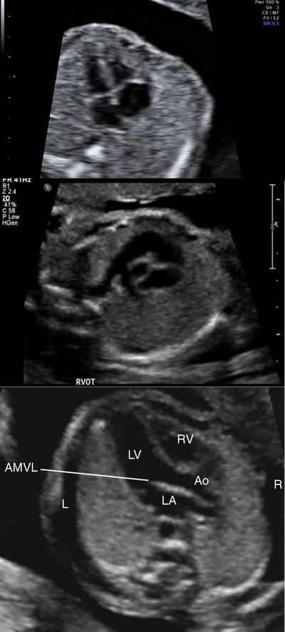
Fetal Echocardiography
Prenatal identification and management of cardiac abnormalities are important since they are the leading cause of infant death and congenital heart disease accounts for 30 to 50% of these deaths.
Fetal echo is recommended for women with family history or previous child born with cardiac anomalies or if there is raised nuchal translucency measurement in the first trimester. Fetal echo is also recommended if other fetal anomalies are seen in routine or anomaly scan.
We at Chennai fetal care include careful assessment of fetal heart as part of the anomaly scan assessment. It’s important to be aware that fetal echocardiography is done to exclude major structural abnormalities in the heart that could alter the outcome of the pregnancy. Minor anatomical variants such as a small hole in the heart muscle (ventricular septal defect) is not always possible to be picked up on routine screening and may only be diagnosed after birth.
Once a complex cardiac abnormality is identified, the couple are counselled regarding the scan findings and the management pathway is discussed. We can also arrange a formal consultation with a specialist paediatric cardiologist / cardiac surgeon if necessary to discuss the management after birth.
Specialised fetal echocardiography would take approximately 30-45 minutes to perform and the scan findings will be discussed with the patient after the scan for further management. A copy of the report will also be sent to the referring Doctor with suggested management plan and follow up.

Fetal echocardiography
Growth and well-being scan with Doppler
This scan is usually offered to pregnant women at later gestation to check for growth and well-being of the fetus. While most obstetricians offer this scan for all their patients, it is particularly useful in high-risk pregnant women (advanced maternal age, IVF pregnancy, pregnancy complicated by preeclampsia, diabetes to name a few) or if there is a suggestion of intrauterine growth restriction (IUGR or FGR) during the pregnancy.
This scan usually takes 15-30 minutes to perform and it involves
1. Measurement of fetal biometry (head, abdomen, femur length) to calculate the estimated fetal weight for the gestation
2. Assessment of placenta and amniotic fluid volume
3. Assessment of fetal movements during the scan
4. Doppler blood flow assessment of placenta and the fetus
The results will be given to the patient after the scan and a copy can be sent to the referring doctor with our recommendations and further follow up.

Fetal Doppler and growth
Multiple pregnancy scan
Multiple pregnancies constitute approximately 1% of all pregnancies. The prevalence of twin pregnancy is around 9-16 per 1000 births in India. In recent times, IVF and assisted conception methods have contributed to the generalised increased of twins and higher-order multiple pregnancies.
Dizygotic twins constitute for approximately 2/3rd of the twin pregnancies with the remaining 1/3rd being monozygotic. The pregnancy-related complications are higher in twins when compared to singletons; in particular, there is an increased risk of preterm delivery and growth restriction (IUGR/FGR) in twins and higher-order multiple pregnancies. In addition, there is a 10-15% risk of developing twin to twin transfusion syndrome (TTTs) in monochorionic twin pregnancies which could result in significant mortality and morbidity to these type of pregnancies.
Multiple pregnancies, therefore, need intense monitoring during the pregnancy when compared to singletons. We at Chennai Fetal Care follow published guidelines and standardised protocols to optimise the management of these pregnancies.
Management for multiple pregnancies in general
1. 12 weeks scan – To establish Chorionicity (Dichorionic or monochorionic) and to perform Down syndrome screening.
2. 20 weeks anomaly scan including cervical assessment.
3. Serial growth scans during pregnancy.
Specific protocol for Dichorionic twin pregnancies
1. In addition to the above, serial growth scans to be performed at 24,28,32 and 36 weeks of gestation with Doppler assessment of placenta and fetus at each visit.
2. Additional intense monitoring if abnormalities are identified during the growth scans.
Specific protocol for Monochorionic twin pregnancies
1. Scans every 2 weeks from 16 weeks until 36 weeks to check for growth, blood flow and specifically to look for signs of twin to twin transfusion syndrome
2. Additional monitoring and option of fetal therapy if complications arise during the gestation
Specific protocol for Monoamniotic twin pregnancies
1. Scans every 2 weeks from 16 weeks until 22 weeks to check for growth, blood flow and specifically to look for cord entanglement. Weekly follow up from 22 weeks upto until 32-34 weeks to prepare for delivery.
2. Additional monitoring and option of fetal therapy if complications arise during the gestation
Higher-order multiple pregnancies
Triplets and quadruplets are relatively rare to occur following natural conception but seen to have a higher incidence following assisted reproductive techniques.
Following counselling for fetal reduction, ultrasound monitoring for fetal well-being is done by serial growth scans every 2 weeks from 16 weeks onwards up to until 32 -34 weeks and delivery is managed according to local policies and guidelines.
The scans for twins usually take around one hour or longer depending upon the complexity of the pregnancy. The scan report is given to the couple after discussing the scan findings. We shall also send a copy of the report to her obstetrician with our suggested management plan and follow up.

Triplet pregnancy